| Interactive Key to the Species of Chaetognatha from the South Atlantic Bight and northern Gulf of Mexico adapted from: Michel, H.B. 1984. Chaetognatha of the Caribbean Sea and adjacent areas. NOAA Technical Reports NMFS 15, United States Department of Commerce. 33 pp. |
|
| How
to use this key: |
Quick Links to Families |
|
Important notes on the classification and morphology of Chaetognatha |
|
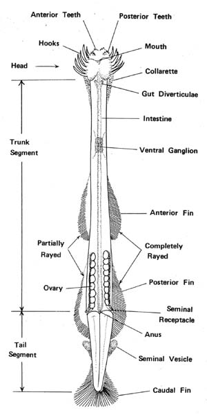
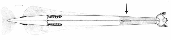
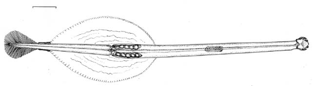
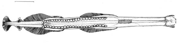
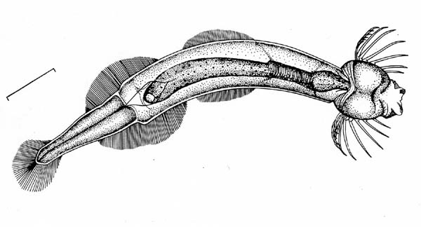
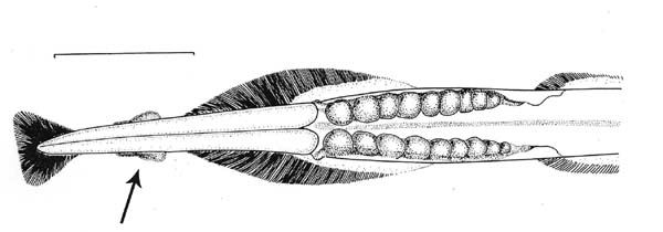
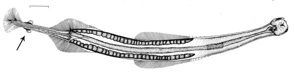
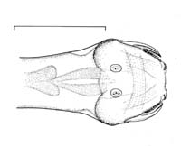
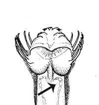
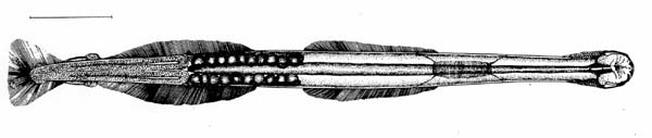
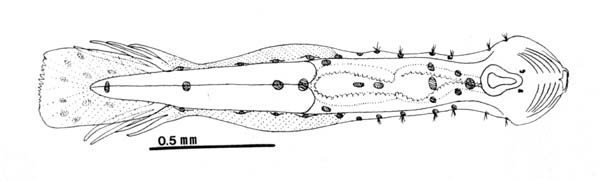
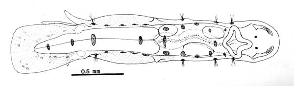
General diagram (bauplan) of a chaetognath
|
|
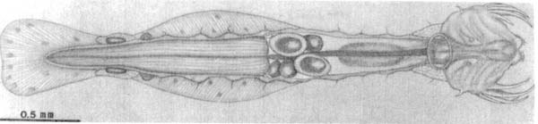
1.a. One pair of lateral fins.....................................................................2 b. Two pairs of lateral fins.................................................................11 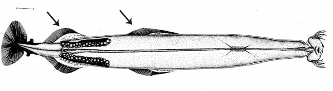
Ventral view of Flaccisagitta enflata (bottom) showing two lateral fins. Scale = 1mm.
|
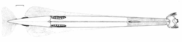
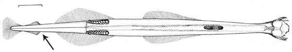
 Ventral view of Pterosagitta draco showing single lateral fin.
Scale = 1 mm
Ventral view of Pterosagitta draco showing single lateral fin.
Scale = 1 mm
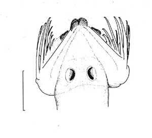
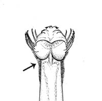
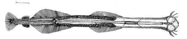
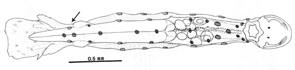
| 2. a. Lateral fins extending onto trunk segment; one set of teeth...........3
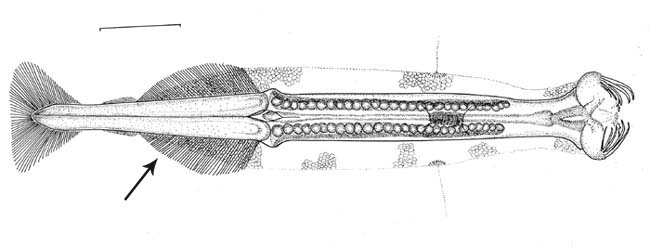
Eukrohnia calliops, ventral view, showing lateral fin extension. Scale = 1 mm. b. Lateral fins limited to tail segment; two sets
of teeth.......................
|

|
|
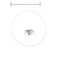
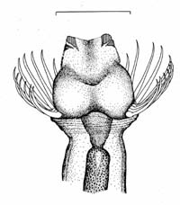
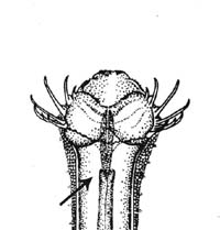
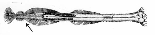
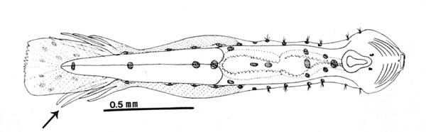
| 4. a. Lateral fins extending to central ganglion - genus Eukronia........5
Eukrohnia
calliops, ventral view, showing lateral fin extension to central
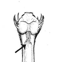
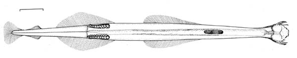
ganglion. Scale = 1.0 mm b. Lateral fins not extending to central ganglion - genus Krohnitta..10 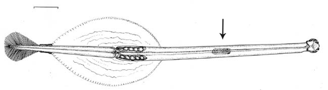 Krohnitta subtilis, ventral
view, showing lateral fin extension posterior to central ganglion.
Scale = 1mm.
Krohnitta subtilis, ventral
view, showing lateral fin extension posterior to central ganglion.
Scale = 1mm. |
|
5. a. Eyes with pigment........................................................................6
b. Eyes without pigment.......................................................................8 |
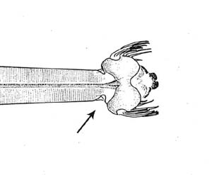
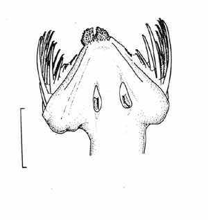
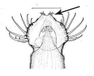
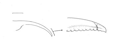
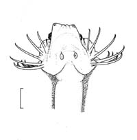
6. a. Apical gland-cell complex bilobate and protruding, causing head to appear pointed; hook tips bent inward at 45-90° angles; transverse musculature extending past posterior edge of ventral ganglion...........7 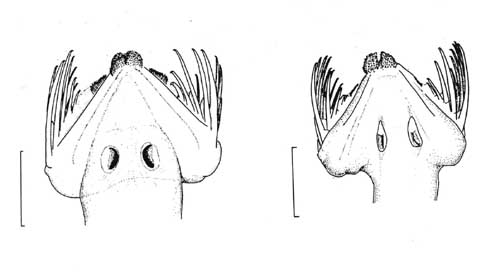
dorsal view of head - Eukrohnia calliops (left); Eukrohnia proboscidea (right). Scale = 0.5 mm
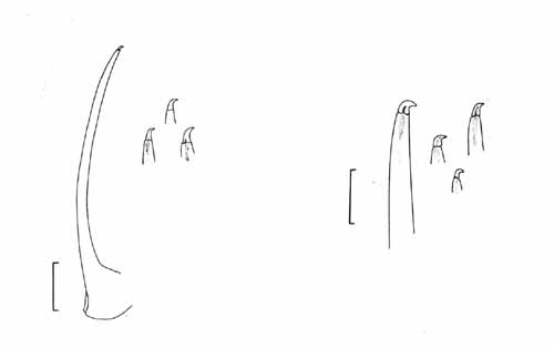
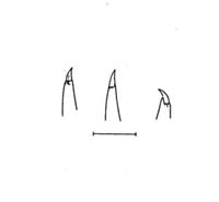
hook tip variations - Eukrohnia calliops (left); Eukrohnia proboscidea (right). Scale = 0.1 mm b. Apical
gland not prominent, a single lobed mass; hook tips straight, transverse
musculature even with posterior edge of ganglion
Eukronia fowleri, immature
specimen, ventral view. Scale = 1 mm
|
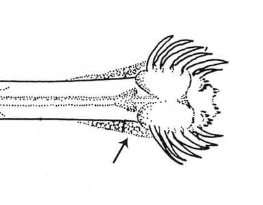
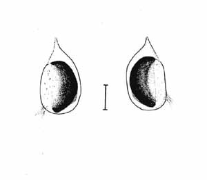
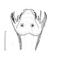
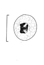
7. a. Eye pigment small, elongate or "U" shaped, in posterior region of eye...............................Eukrohnia proboscidea Furnestin and Ducret, 1965
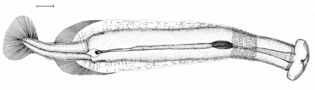
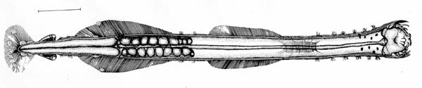
Eukrohnia proboscidea (top) whole animal, dorsal view; head, (bottom left) dorsal view (scale = 0.5 mm); (bottom right) left eye (scale = 0.1 mm). b. Eye pigment large, lunate, encompassing most of median portion of eye..............................................................Eukrohnia calliops
Eukrohnia calliops, whole specimen, ventral view. Scale = 1 mm.
Eukrohnia
calliops head, dorsal view (left)
(scale = 0.5mm); eyes (right) (scale = 0.1 mm) |
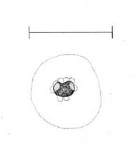
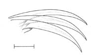
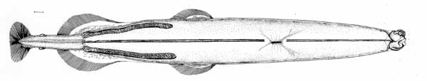
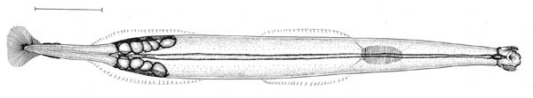
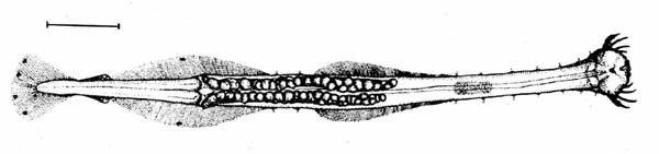
8. a. Number of hooks greater than 11, hook tips straight...................... .........................................................Eukrohnia bathyantarctica David ,1958
Eukrohnia bathyantarctica, whole animal,
dorsal view. b. Number of hooks less than 11, hook tips bent inward........................9
|
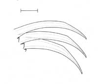
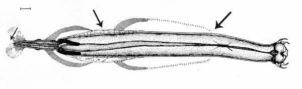
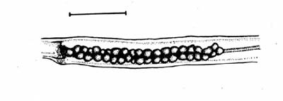
9. a. Hooks stout, nearly straight; tail less than 25% of body length........ ...............................................................Eukrohnia hamata (Mobius, 1875)
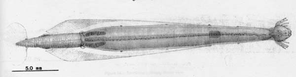
Eukrohnia hamata whole specimen, dorsal view.
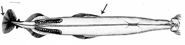
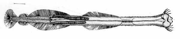
b. Hooks slender, gently curved; tail usually greater than 25% of body length ..............................................Eukrohnia bathypelagica Alvarino, 1962
Eukrohnia bathypelagica, whole animal, dorsal view.
|
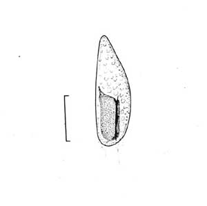
10.a. Outer margins of hooks obtusely
angled; mature ovaries elongate, may extend past edge of lateral
fins............Krohnitta pacifica (Aida,
1897)
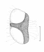
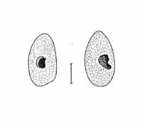
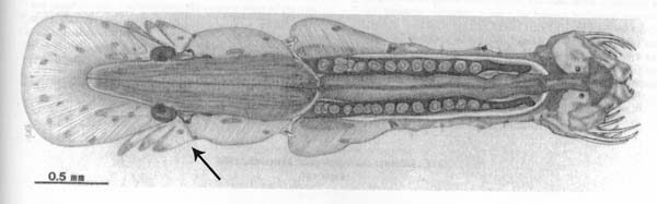
Krohnitta pacifica: (top) whole animal, ventral view (scale = 1mm); (bottom left) right eye (scale = 0.05 mm); (bottom right) hooks (scale = 0.1 mm). 10b. Outer margin of hooks evenly rounded; mature ovaries compact, not extending past edge of lateral fins.........Krohnitta subtilis (Grassi, 1881)
Krohnitta subtilis: (top) whole animal, ventral view (scale = 1mm); (bottom left) right eye (scale = 0.05 mm); (bottom right) hooks (scale = 0.1 mm).
|
|
(this genus is referred to as Sagitta in Michel, 1984 and Casanova, 1999; as Flaccisagitta in McLelland 1989 and Bieri, 1991) b. Body rigid, translucent; trunk musculature prominent......................14 |
12a. Anterior fins long, inserted close
to ventral ganglion, connected to posterior fins by raised portion of
body cuticle; caudal fin bilobate (this genus is referred to as Sagitta in Michel, 1984 and Casanova, 1999; as Flaccisagitta in McLelland 1989; as Pseudosagitta in Bieri, 1991)
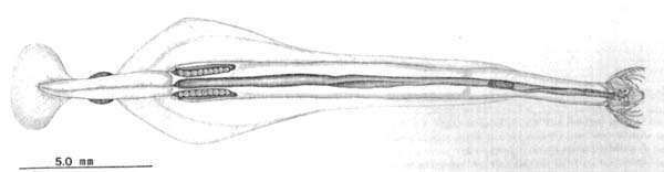
Ventral views of Flaccisagitta lyra whole animal (top) and head (bottom). Scale = 1mm. Flaccisagitta enflata, ventral view. Scale = 1 mm.
|

13.a. Two to four elongate, thin anterior teeth which sometimes protrude anteriorly .........................Flaccisagitta hexaptera (d'Orbigny, 1843) (this genus is referred to as Sagitta in Michel, 1984 and Casanova, 1999; as Flaccisagitta in McLelland 1989 and Bieri, 1991)
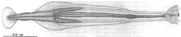
Flaccisagitta hexaptera whole animal (top), ventral view; head (bottom left) and anterior teeth, dorsal view (scales = 1 mm); right eye (bottom right) (scale = 0.015 mm) b. Four to eight short, wide, overlapping anterior teeth............................ .................................................................Flaccisagitta enflata (Grassi, 1881) (this genus is referred to as Sagitta in Michel, 1984 and Casanova, 1999; as Flaccisagitta in McLelland, 1989 and Bieri, 1991)
Flaccisagitta enflata whole animal (top), ventral view (scale = 1 mm); right eye pigments (bottom)(scale = 0.015 mm).
|
|
14. a. Hooks finely serrate on inner margins as seen under 100x magnification ........................... Serratosagitta serratodentata (Krohn, 1853) (this genus is referred to as Sagitta in Michel, 1984 and Casanova, 1999; as Serratosagitta in McLelland, 1989 and Bieri, 1991)
Serratosagitta serratodentata
whole animal, ventral view (top)(scale = 1 mm); b. hooks not serrate..............................................................................15
|
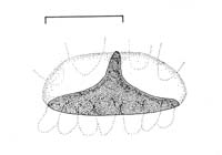
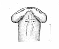
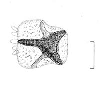
15. a. Collarette absent or indistinct.....................................................16 Caecosagitta macrocephala head, ventral view, showing lack of collarette. Scale = 1.0 mm. Solidosagitta planctonis head, ventral view, showing distinct collarette.
|
16. a. Eyes without pigment; anterior fins entirely
rayed, emerging at a point well separated from ventral ganglion (this genus is referred to as Sagitta in Michel, 1984 and Casanova, 1999; as Caecosagitta in McLelland, 1989 and Bieri, 1991)
Caecosagitta macrocephala (top) immature specimen, ventral view; (bottom) head, ventral view. Scales = 1 mm.
|
17. a. Tail segment less than 22% of total body length; mature ovaries compact, with three to five large ova......... Mesosagitta minima (Grassi, 1881) (this genus is referred to as Sagitta in Michel, 1984 and Casanova, 1999; as Mesosagitta in McLelland, 1989 and Bieri, 1991)
Mesosagitta minima, ventral view. Scale = 1 mm.
|
18. a. Seminal vesicles located approximately equal distance from posterior fins and caudal fin; maximum body length at maturity less than 15 mm ..........................................Mesosagitta decipiens (Fowler ,1905) (this genus is referred to as Sagitta in Michel, 1984 and Casanova, 1999; as Mesosagitta in McLelland, 1989 and Bieri, 1991)
Mesosagitta decipiens posterior end of body, ventral view, showing position of seminal vesicles. Scale = 1 mm.
b. Seminal vesicles adjacent to caudal
fin and well separated from posterior fins; maximum body length at maturity
may reach 20mm (this genus is referred to as Sagitta in Michel, 1984 and Casanova, 1999; as Mesosagitta in McLelland, 1989 and Bieri, 1991)
Mesosagitta sibogae (top) whole animal, ventral view; (bottom) head, dorsal view. Scales = 1 mm.
|
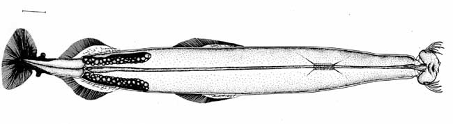
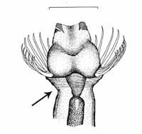
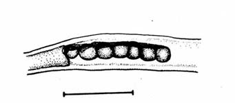
19.a. Gut diverticulae present* ............................................................20 Solidosagitta planctonis (left) and Ferosagitta hispida (right) showing presence of gut diverticulae. * gut diverticulae are often undeveloped and difficult to see in immature specimens. b. Gut diverticulae absent................................genus Sagitta..........21 Sagitta megalophthalma showing lack of gut diverticulae.
|
| 20.a. Posterior fins triangular with
prominent rayless zone; walls of gut tube lined with large vacuolar
cells (this genus is referred to as Sagitta in Michel, 1984 and Casanova, 1999; as Solidosagitta in McLelland, 1989 and Bieri, 1991)
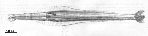
Solidisagitta planctonis (top) whole animal, ventral view (scale = 1 mm); (bottom left) head, dorsal view (scale = 0.5 mm); (bottom right) right eye (scale = 0.1 mm). b. Posterior fins rounded, completely rayed; no large vacuolar cells associated with gut tube ..........................Ferosagitta hispida (Conant, 1895) (this genus is referred to as Sagitta in Michel, 1984 and Casanova, 1999; as Ferosagitta in McLelland, 1989 and Bieri, 1991)
Ferosagitta hispida whole animal, ventral view. Scale = 1 mm.
|

21.a. Posterior fins well separated from seminal vesicles....................22
Sagitta megalophthalma whole animal, ventral view, showing position of seminal vesicles. Scale = 1 mm. b. Posterior fins contacing the seminal vesicles....................................23
Sagitta helenae, whole animal, ventral view, showing position of seminal vesicles. Scale = 1 mm.
|
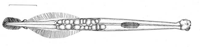
22. a. Anterior fins emerging posterior to ventral ganglion by a distance of about half the length of the ganglion; large vacuolar cells lining middle third of gut tube............Sagitta megalophthalma Dallot and Ducret, 1969 (this genus is referred to as Sagitta in Michel, 1984 and Casanova, 1999; as Sagitta in McLelland, 1989; as Parasagitta in Bieri, 1991)
Sagitta megalophthalma (top) whole animal, dorsal view (scale = 1 mm); (bottom left) head, dorsal view (scale = 0.5 mm); (bottom right) left eye (scale = 0.1 mm) b. Anterior fins emerging level with posterior
edge of ventral ganglion; no large vacuolar cells associated with gut
tube
Sagitta bipunctata, whole animal, ventral view. Scale = 1 mm.
|
23. a. Anterior teeth numerous (8-18), elongate, protruding
outward in an overlapping, fan-shaped arrangement
Sagitta helenae (top) whole animal, ventral view; (bottom left) head, dorsal view; (bottom right) anterior head, ventral view. Scales = 1 mm. b. Anterior teeth less numerous (6-9), short, lying flat against head....24
|

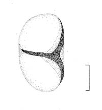
24. a. Mature ovaries with large ova which are few in number and arranged in one row.............................................Sagitta tenuis Conant, 1896 (this genus is referred to as Sagitta in Michel, 1984, Casanova, 1999 and McLelland, 1989; as Parasagitta in Bieri, 1991)
Sagitta tenuis (top) whole animal, ventral view; (bottom) lateral view of ovaries. Scales = 1 mm. b. Mature ovaries with small, numerous ova arranged in double rows ........................................................Sagitta friderici Ritter-Zahony, 1911 (this genus is referred to as Sagitta in Michel, 1984, Casanova, 1999 and McLelland, 1989; as Parasagitta in Bieri, 1991)
Sagitta friderici (top) whole animal, ventral view; (bottom) lateral view of ovaries. Scales = 1 mm.
|
|
Paraspadella schizoptera, dorsal view, showing digitate tail structures.
b. Without adhesive structures but with adhesive glands on the ventral surface of the tail......................... Spadella cephaloptera (Busch, 1851)
Spadella cephaloptera, dorsal view.
|
26. a. Adhesive structures originate from the posterior edge of the seminal vesicles, are attached laterally, and are confluent with the caudal fin ............................................Paraspadella pulchella Owre, 1963 (this genus is referred to as Spadella in Michel, 1984)
Spadella puchella whole animal, dorsal view, showing position of adhesive structures.
Paraspadella schizoptera, whole animal, dorsal view, showing position of adhesive structures.
|
27. a. Each adhesive structure is divided into four to six slender processes .........................................Paraspadella schizoptera Conant, 1895 (this genus is referred to as Spadella in Michel, 1984)
Spadella schizoptera, whole animal, dorsal view.
b. Each adhesive structure is divided into
two slender processes, or, in very small specimens, the structure may
be undivided (this genus is referred to as Spadella in Michel, 1984)
Spadella nana, whole animal, dorsal view.
|
The Classification of the Chaetognatha This interactive key is an adaptation of the work of McLelland (1989) and Michel (1984) and covers the species known from the northwestern Atlantic. Since the publication of these papers, there have been many disagreements amongst workers regarding the classification of the genera within the Chaetognatha. However, without a cladistic examination of this group, the classification of its species and genera remains uncertain. For the purposes of this guide we follow the classification used by McLelland (1989) and Michel (1984) (as updated by Salvini-Plawen, 1986) At each terminal couplet, we include notes on the different terminologies used by more recent authors. The family Sagittidae has been subjected to tremendous revision during the last several decades as a result of the morphological grouping of species within this family. Tokioka (1965) proposed that the genus Sagitta be broken down into 9 smaller genera (seven of which are used in this key: Serratosagitta, Parasagitta, Ferosagitta, Mesosagitta, Solidosagitta, Caecosagitta, and Flaccisagitta). His revision was not retained in his later work but was used by McLelland (1989) and in a revised form by Bieri (1991). In his review of south Atlantic chaetognaths, Casanova (1999) argued against the use of Tokioko's revision based on what he perceived as a lack of cladistical support for the genera as well as the high degree of morphological variability seen in juveniles. Please refer to Bieri (1991) and Casanova (1999) for more rigorous reviews of the taxonomy of the family Sagittidae. The family Spadellidae was recently divided into multiple genera. Species of Spadella were included within the Caribbean key to chaetognaths (Michel, 1984) but were absent from McLelland’s (1989) Gulf of Mexico review. They are included here because it is likely that their distribution may extend into South Atlantic Bight epibenthic and benthic communities. The appropriate taxonomic changes have been made to this key to accommodate the updated classifications erected by Salvini-Plawen (1986) and Bowman and Bieri (1989), however it is important to note that the due to the benthic nature of some spadellids, their distributions and morphological variability are poorly known. The classification of Eukrohnia, Krohnitta and Pterosagitta
species used in this key has remained static in past and current literature.
Classification of the Chaetognatha of the SAB and Gulf of Mexico Phylum Chaetognatha (Leukardt, 1894)
Chaetognaths exhibit a high degree of morphological variation as a function of their maturity and environment. This aspect has, in part, caused the classification of the Chaetognatha to be currently in a state of flux. It is important to note some of the more common variants for identification purposes (from Bieri 1991): size of mature individuals References: Bieri R. (1991) Systematics of the Chaetognatha. In: The Biology of Chaetognaths. Q Bone, H Knapp, AC Pierrot-Bults, eds. Oxford Scientific Publishing. pp 122-136. Bowman, TE and Bieri, R (1989) Paraspadella anops, new species, from Sagittarius Cave Grand Bahama Island, the second troglobitic chaetognath. Proceedings of the Biological Society of Washington 102(3):586-589. Casanova, J. (1999) Chatognatha. In: South Atlantic Zooplankton. D. Boltovskoy, ed. Backhuys Publishers. pp. 1353-1374. McLelland, JA. (1989) An illustrated key to the Chaetognatha of the northern Gulf of Mexico with notes on their distribution. Gulf Research Reports 8(2):145-172. Michel, HA. (1984) Chaetognatha of the Caribbean Sea and adjacent areas. NOAA Technical Report NMFS 15. US Department of Commerce. 33 p. Salvini-Plawen, LC. (1986). Systematic notes on Spadella and on the Chaetognatha in general. Zeitschrift fur zoologische Systematik und Evolutionsforschung 24(2): 122-128. Tokioka, T. (1965) The taxonomical outline of Chaetognatha. Publications of the Seto Marine Biological Laboratory 12:335-357.
Please cite page as: Southeastern Regional Taxonomic Center [SERTC]. 2004 December. Chaetognaths
of the South Atlantic Bight and the Northern Gulf of Mexico. SERTC Taxonomic
Information and Educational Resources web page. <https://www.dnr.sc.gov/marine/sertc/Chaetognath%20key/Chaetognath%20key.htm>.
Accessed Yr Mon da.
|