CONTENTS
Introduction
The South Atlantic Bight
Methods
Octocoral Morphology
Glossary
Gorgonacean
Bauplan
see this for keys
Notes on the Species
Carijoa
riisei
Scleranthelia
rugosa
Telesto fruticulosa
Telesto nelleae
Telesto sanguinea
Bellonella rubistella
Pseudodrifa nigra
Nidalia occidentalis
Iciligorgia schrammi
Diodogorgia
nodulifera
Titanideum
frauenfeldii
Muricea pendula
Thesea nivea
Bebryce cinerea
Bebryce parastellata
Scleracis guadalupensis
Paramuricea sp.
Leptogorgia hebes
Leptogorgia punicea
Leptogorgia
cardinalis
Leptogorgia virgulata
Leptogorgia setacea
Leptogorgia euryale
Viminella
barbadensis
Renilla reniformis
Sclerobelemnon
theseus
Stylatula elegans
Virgularia presbytes
| Guide
to the Shallow Water (0-200 m) Octocorals of the South Atlantic
Bight.
S. T. DeVictor
& S. L. Morton, 2007
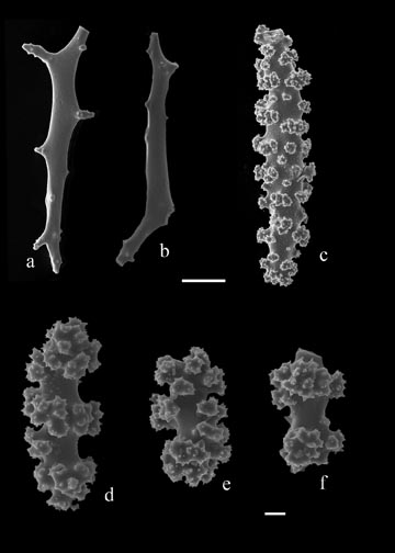
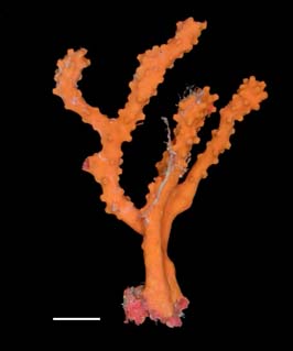
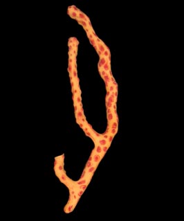
Diodogorgia nodulifera
(Hargitt, in Hargitt and Rogers, 1901) Remarks. Fragments of the only two samples of this species recorded in the SAB were examined for this work, as well as a sample from a colony (USNM 49705) collected south of the SAB for comparison because of the variance in colony morphology. The southern colony displays the most common form and could conceivably be found in the SAB. The southern colony sample is yellow with red, moderately protruding polyp mounds and the cylindrical stem is approximately 5 mm in diameter. A ring of boundary canals divides the cortex and medulla. The cortex has a dense outer layer and spongy inner layer separated by a plexus of solenia. The outer cortex and polyp mounds contain small tuberculated spheroids, tuberculated, irregularly branched bodies, capstans and slender, warty, amber spindles. Also present are elongated, sparsely warted spindles. The neck of the polyps contain small, red tuberculated spheroids and branched bodies. The medulla contains light pink warted rods that are occasionally branched. Atlantic distribution: Georgia to Surinam, Gulf of Mexico, Caribbean, 20-183 m (Deichmann, 1936; Bayer, 1959; Bayer, 1961; NMNH collections; SERTC collection)
|
Figure 3. Sclerites of Diodogorgia nodulifera (S2698). a,b) rods of medulla; c) spindle of cortex; d-f) radiates of cortex. Scale bar for a-c = 50 µm; d-f = 10 µm. |